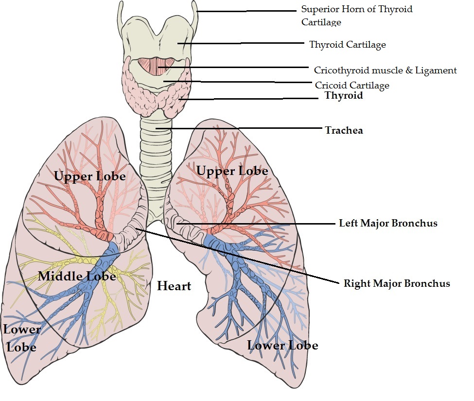
Trachea
Definition
Tube that connects the pharynx and larynx to the lungs, allowing the passage of air
The Extent
Extends from the larynx and branches into the two primary bronchi.
Situation
Trachea lies in the midline of the neck
Below the Cricoid cartilage
The tracheal bifurcation is at the level of sternal angle (T5)
Structure
Below the larynx lies the cricoid cartilage
Crico-tracheal ligament lies between the lower border of cricoid and the upper end of trachea.
Trachea has 18 - 22 C shaped cartilagenous rings
Annular Ligament - between two rings
Trachealis muscle connects the ends of the incomplete rings; ovrlies oesophageal muscle and forms the posterior wall of trachea
Inner surface pinkish
Stratified ciliated columnar epithelium lines the inner surface; the epithelium contains mucous glands and musle fibres.
Longitudinal elastic fibers enable the trachea to stretch and descend with the roots of the lungs during inspiration.
The trachea divides between the T5 and T7 vertebral levels.
The carina - a ridge seen internally at the bifurcation and is a landmark during bronchoscopy.
The arch of the aorta is at first anterior to the trachea and then on its left side immediately superior to the left main bronchus.
Other close relations include the brachiocephalic and left common carotid arteries.
Blood Supply
The inferior thyroid arteries.
Nerve Supply
Its smooth muscle is supplied by parasympathetic and sympathetic fibers - by the vagi.
Applied Anatomy
Trachiostomy - done to reduce respiratory effort
Bronchi
Main bronchi
Right and Left
Each main bronchus extends from the tracheal bifurcation to the hilus of the' corresponding lung.
The right main bronchus - has
(1) an upper part, from which the segmental bronchi for the upper lobe arise
(2) a lower part, from which the segmental bronchi for the middle and lower lobes arise
The left main bronchus divides into two
(1) one for the upper lobe
(2) one for lower lobe.
The upper lobar bronchus has an upper division and a lower, or lingular, division.
The right main bronchus shorter, wider, and more vertical than the left.
So foreign objects traversing the trachea are more likely to enter the right main bronchus.
The left main bronchus crosses anterior to the esophagus, which it indents.
Both bronchi have cartilaginous rings that are replaced by separated plates at the roots of the lungs.
Blood Supply
The bronchi supplied by the bronchial arteries and veins,
Nerve Supply
Its smooth muscle is supplied by parasympathetic and sympathetic fibers - by the vagi.


