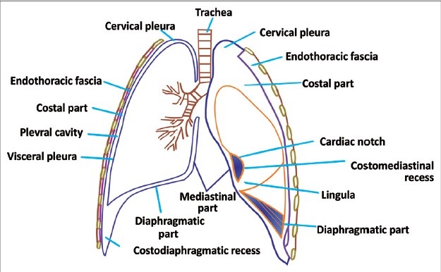
The Pleura
Definition
The membrane lining a body cavity containing the lungs and covering the lung also
The lungs are surrounded by two serous membranes, the pleurae.
The layers
The outer pleura (parietal pleura) is attached to the chest wall.
The inner pleura (visceral pleura) covers and is attached to the lung and
other structures, i.e. blood vessels, bronchi and nerves.
The pleura is derived from embryonic coelomic lining
Between the two is a thin space known as the pleural space
The space contains a small amount of pleural fluid.
The parietal pleura is highly sensitive to pain; the visceral pleura is not, because
it receives no nerves of general sensation.
The various parts
of the parietal pleura are :
costal
diaphragmatic
mediastinal
cervical (cupola)
The surfaces and cavities
In health they are in actual contact with one another
But the potential space between them is known as the pleural cavity.
The adjacent surfaces of the pleura are moistened by a serous fluid
When the lung collapses or when air or fluid collects between the two layers, the cavity becomes apparent.
The right and left pleural sacs are entirely separate from one another;
between them are all the thoracic viscera except the lung.
on the sides the lung does not fill the sac formed by the two layers of the pleura - costophrenic sinus -
in the chest X-ray - costophrenic angle
The right pleural sac is shorter, wider, and reaches higher in the neck than the left.
Pulmonary Ligament : the root of the lung is covered Here the pleura forms
a fold called the pulmonary ligament,
Structure of Pleura :
the pleura is covered by a single layer of flattened, nucleated cells
Vessels and Nerves.—The arteries of the pleura are derived from the intercostal,
internal mammary, musculophrenic, thymic, pericardiac, and bronchial vessels.
The veins correspond to the arteries.
The nerves are derived from the phrenic and sympathetic nerves along the vessels


