Brain and its Parts
Parts of the brain
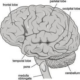
The cerebrum
Cerebellum
Pons varolii
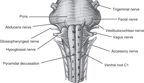
Medulla oblongata
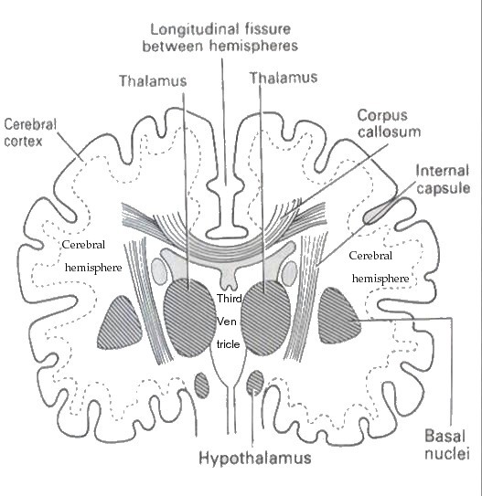
Thalamus
Internal capsule
Basal nuclei
Hypothalamus
Midbrain
Covering of the brain
Duramater
Arachnoid mater
piamater
The Cerebrum
Lobes
frontal
parietal
temporal
occipital
The ridges in the cerebrum are called the gyri (gyrus)
The grooves are called the sulci (sulcus)
The ridge in front of the central sulcus is called the precentral gyrus
The one behind the central sulcus is called the post-central gyrus
The Interior
Cerebral cortex is composed of nerve cell bodies on the surface - grey matter
Inside the cerebrum are the nerve fibres (tracts) which connect the various lobes - white matter
The afferent and efferent fibres linking the different parts of the brain and spinal cord are:-
Arcuate fibres - interconnecting fibres between the different lobes of the same hemispheres
Commisural fibres - connecting one hemisphere to the other e.g. corpus callosum
Projection fibres - which connect the cerebral cortex to grey matter of lower parts of the brain and the al cord e.g. the internal capsule
Functions and Functional Areas of Cerebrum
Precentral (motor) area - initiates the contraction of the voluntary muscles
Premotor area - manual dexterity
Lower part of premotor area - speech (motor) (Broca's area)
Frontal area or pole - behaviour, character, intellectual trait
Postcentral area (sensory) - pain, temperature, pressure, touch, knowledge of muscular elements and the position of joints
Parietal area - knowledge of objects (e.g.touch and recognise)
Sensory speech area - situated in the lower part of the parietal lobe and part of the temporal - perception of speech
Thalamus
The thalamus consists of two masses of nerve cells and fibres situated within the cerebral hemispheres just below the corpus callosum one on each side of the third ventricle
This region contains a cluster of nuclei
Most of the sensory inputs are conducted to the cerebral cortex through the thalamus (spinothalamic tracts)
Axons carrying auditory, visual and other sensory informations synapse with specific nuclei of region
This region may also influence mood and general body movements due to strong emotions such ar or anger
Hypothalamus
Hypothalamus - The master control of the autonomic nervous system, parasympathetic and sympathetic. This system stimulates and controls structures such as the heart, most glands and smooth muscles. In effect, this system allows your systems to excite and relax, as needed. This system integrates autonomic and endocrine functions with behavior.
Hypothalamus
This region contains small nuclei and nerve tracts
The nuclei called mammillary bodies are involved in olfactory reflexes and emotional response ours
The funnel shaped infundibulu from the hypothalamus connects to the posterior pituitary or ohypophysis
This region controls the secretions of pituitary gland
The hypothalamus receives inputs from several sensory systems such as tongue,nose and rnal genitalia.
It is associatedwith emotional and mood relationships
It provides a relaxed feeling
A section of the cerebrum showing some connecting nerve fibres
Basal Ganglia
This area of grey matter lies deep within the cerebral hemispheres
Consist of the caudate nucleus, putamen and globus pallidus.
They are functionally important for controlling voluntary movements and establishing postures.
It influences skeletal muscle tone
When they are altered - say in disorders like Huntington disease or Wilson disease - the person unwanted movements, such as involuntary jerking movements of an arm or leg or spasmodic ement of facial muscles.
The caudate nucleus and putamen along with the interposed anterior limb of the internal capsule are collectively known as the corpus striatum (i.e. striated body) because of their appearance.
Similarly, the shape of the putamen and globus pallidus resembles a lens, and they are collectively called the lenticular nucleus.
Internal Capsule
Consists of projection fibres - fibres connecting the cerebral cortex with the lower parts of the brain and the spinal cord
Lies deep within the brain
Between the basal ganglia and the thalamus
All the impulses passing to and from the cerebral cortex are carried by the fibres in the internal capsule
The motor fibres within the internal capsule form the pyramidal tracts (otherwise called the corticospinal tracts)
Pyramidal fibres cross to the other side at the level of the medulla oblongata (called decussation)
Midbrain
Situated around the cerebral aqueduct between the cerebrum and pons varolii
Consists of nerve cells and nerve fibres connecting the cerebrum with lower parts of the brain with the spinal cord
The nerve cells serve as relay stations for the ascending and descending fibres
The roof of this region contains four nuclei
The nuclei form mounds
They are collectively called corpora quadrigemina
It is formed of two superior colliculi and two inferior
The superior colliculi are involved in visual reflexes
They control eye and head movements
They aid in visual tracking of moving objects
The inferior colliculi are involved in hearing
Cerebellum
An organ concerned with the co-ordination of the voluntary movements
Maintains posture and balance
Situated behind pons varolii
Below the posterior portion of the cerebrum
Sits in the posterior cranial fossa
Ovoid in shape with two hemispheres
There is a median strip of tissue called vermis
Grey mater forms outer surface
White mater inside
Cerebellum communicates with other regions of CNS through three large nerve tracts called the cerebellar peduncles
Consitst of the following three parts :
Sensory input for cerebellum derived from
Muscles and joints
Eyes
Ears (from semicircular canals)
Applied Anatomy
Cerebe;;ar damage dut to injury or infarction may cause
Decreased muscle tone
Imbalance
Lack of co-ordination
Medulla Oblongata
Extends from the pons varolii above and is continuous with the spinal cord below
2.5 cm
Situated within the cranium above the foramen magnum
Central fissures anteriorly and posteriorly
Vital centres
Medulla oblongata contains vital centres
Cardiac centre
Respiratory centre
Vasomotor centre
Reflex centres of vomiting, coughing, sneezing and swallowing
Special features
Decussation of the pyramids - motor fibres from motor area in the pyramidal tracts cross
Sensory decussation - some of the sensory nerves ascending from the spinal cord cross - others cross in the spinal cord itself
Functions
Cardiac centre - controls the rate and force of cardiac contraction sympathetic and parasympathetic nerve fibres originating in the medulla pass to the heart. Sympathetic stimulation increases the rate and force of heart beat - parasympathetic does the opposite
Respiratory centre : controls the rate and depth of respiration. Impulses pass to the phrenic and intercostal nerves.
Gets stimulated by excess of CO2 and to a lesser extent, by deficiency of O2 by nerve impulses from the chemoreceptors from the carotid bodies
Vasomotor centre : controls the diameter of the blood vessels : small arteries and arterioles through the autonomic nervous system
Reflex centres : irritating substances in the stomach or respiratory tract - stimulate reflex centres - reflex actions of vomiting coughing and sneezing.
Reticular Formation
A collection of neurones in the core of the brain stem
constanttly receives "information" being transmitted in ascending and descending tracts
Surrounded by neural pathways which pass nerve impulses between the brain and the spinal cord
It has synaptic links with other parts of the brain
Functions
of reticular formation
Coordination of skeletal muscle activity associated with voluntary motor movement and the maintenance of balance
Coordination of activity controlled by the autonomic nervous system, e.g. cardiovascular, respiratory and gastrointestinal activity
Selective awareness that functions through the reticular activating system which selectively blocks or passes sensory information to the certebral cortex
Reticular formation
Reticular activating system
Cell Bodies
Groups of cell bodies are called nuclei in the central nervous system and ganglia in the peripheral nervous system
The medulla oblongata (or medulla) is located in the hindbrain, anterior to the cerebellum. It is a cone-shaped neuronal mass responsible for autonomic (involuntary) functions ranging from vomiting to sneezing. The medulla contains the cardiac, respiratory, vomiting and vasomotor centers and therefore deals with the autonomic functions of breathing, heart rate and blood pressure.
The bulb is an archaic term for the medulla oblongata and in modern clinical usage the word bulbar (as in bulbar palsy) is retained for terms that relate to the medulla oblongata, particularly in reference to medical conditions. The word bulbar can refer to the nerves and tracts connected to the medulla, and also by association to those muscles innervated, such as those of the tongue, pharynx and larynx.


