The Testis
Definition
reproductive glands of the male
4.5 cm x 2.5 cm x 3 cm
Suspended in the scrotum by the spermatic cords
Embryology
Develop in the lumbar region of the abdominal cavity just below he kidneys
descend into the scrotum taking with them coverings of peritoeum, blood and lymph vessels, nerves and the deferent duct
the coverings get detached from the abdominal peritoneum
descent complete by the 8th month of fetal life.
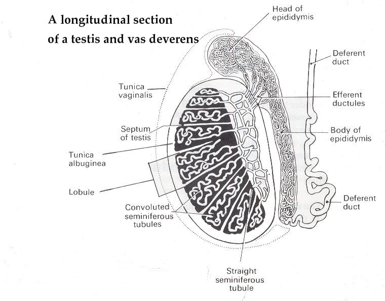
Structure
surrounded by three layers of tissue
The tunica vaginalis - outer covering - a downgrowth of the abdominal and pelvic peritoneum
Tunica albuginea - a fibrous covering surrounding the testes -deeper to tunica vaginalis
ingrowths from tunica albuginea form septa dividng the glandular structure of the testes into lobules
Tunica vasculosa - a network of capillaries supported by delicate connective tissu
Structure of the testes
200 to 300 lobules
within lobule 1 to 4 convoluted loops composed of germinal epitelial cells called seminiferous tubules.
Between the tubules groups of interstitial cells (of Leydig) - secrete testosterone after puberty
at the upper pole of the testis the tubules combne to form a single tortuous tubule, the epdidymis
epdidymis leaves the scrotum as the deferent duct (vas deferens) in the spermatic cord.
blood and lymph vessels pass to the testes in the spermatic cords
Spermatic cords
Two rope like structures by which the testes is hung in the scrotal sac
2 spermatic cords one for each testis
consists of
1 testicular artery
1 testicular venous plexus
lymph vessels
1 deferent duct (vas deferens)
nerves
Within a sheath of fibrous and connective tissue and smoth muscle.
passes through the inguinal canal.
at the deep inguinal ring the structures within the cord diverge
The testicular artery from the abdominal aorta just below the renal arteries
The testicular vein passes into the abdominal cavity
the left vein opens into the left renal vein and the right into the inferior vena cava.
The lymph drainage is through lymph nodes around the aorta.
The deferent duct is 45 cm long
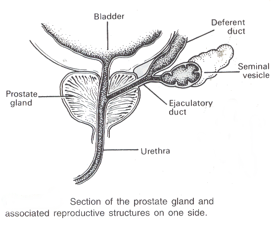
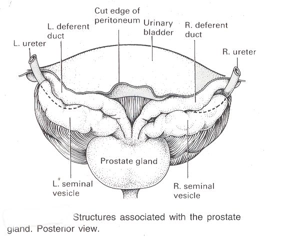
passes upwards from the testis through the inguinal canal and ascends medially towards the posterior wall of the bladder where it is joined by the duct from the seminal to form the ejaculatory duct
The nerve supply is provided by branches from the 10th amd 11th thoracic nerves.
Seminal vesicles are two small fibromuscular pouches line with columnar epithelium, lying on the posterior aspect of the bladder
Seminal vesicles open into a short duct which joins with the corresponding deferent duct to form an ejaculatory duct.
They secrete viscous fluid that helps to keep the spermatozoa alive
Ejaculatory ducts
two tubes
2 cm long
each formed by the union of the duct from a seminal vesicle and a deferent duct.
They pass through the prostate gland and join the prostatic urethra, carrying seminal fluid and spermatozoa to the urethra