The Spleen
Definition
The largest lymph organ in the body
Formed by reticular and lymphatic tissue
Situation
Left hypochondrium
Abdomial cavity
Between the fundus of the stomach and the diaphragm
Purple
12 x 6 x 2 cm
200g
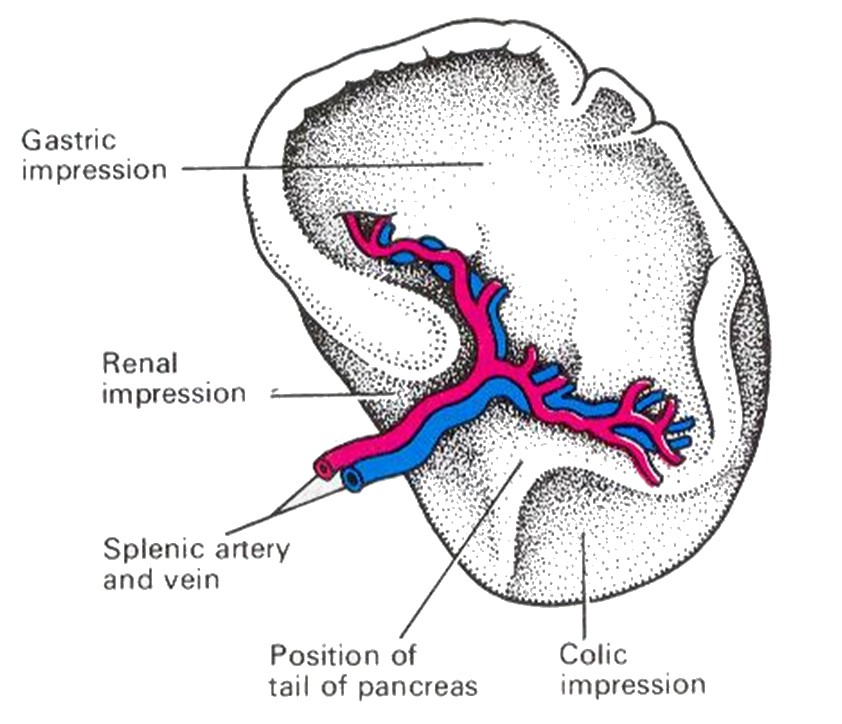
Organs associated with the spleen
Superiorly and posteriorly _ diaphragm
Inferiorly - left colic flexure of the large intestine
Anteriorly - fundus of stomach
Medially - pancreas and the left kidney
Laterally - separated from the 9th , 10th and 11th ribs and the intercostal muscles by the diaphragm
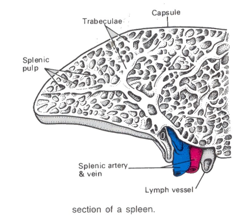
Structure
Oval
Hilum on the lower medial border
Anterior surface covered by peritoneum
Fibroelastic capsule
The capsule dips into the spleen forming trabeculae
Splenic pulp - red pulp and white pulp
Red pulp filled with blood
White pulp consists of areas of lymphatic tissue and blood vessels
Lymphocytes and macrophages around blood vessels
Hilum
The structures entering and leaving the spleen at the hilum are :
Splenic artery
Splenic vein
Lymph vessels efferent only
Functions
blood passing through the spleen flows in sinuses which have distinct pores between the endothelial cells, allowing it to come into close association with splenic pulp
Phagocytosis
Old and abnormal erythrocytes are destroyed in the spleen and the breakdown products, bilirubin and iron , are passed to the liver via the splenic and portal veins
Other cellular material, e.g. Leukocytes, platelets and microbes, are phagocytosed in the spleen
No afferent lymphatics
Development of lymphocytes
B and T lymphocytes multiply and become immunocompetent in the spleen
Some B lymphocytes develop into plasma cells and produce antibodies
Erythropoiesis production of RBCs
Applied Anatomy
Hypersplenism - spherocytosis - both cause excessive destruction of RBCs → Haemolytic jaundice - needs splenectomy